You can only learn so much about exactly how a building was compromised and eventually fell by looking at the pile of rubble left behind to know how to prevent it from falling next time. Such is the case with the debilitating condition cervical stenotic myelopathy (CSM), commonly known as Wobbler syndrome—a foal that’s grown too fast or suffered some kind of physical trauma, among other factors, exhibits a stack of debilitating neurologic signs. Researchers still have much to learn about why this disease develops and how to best treat affected horses.
Jennifer Janes, DVM, PhD, Dipl. ACVP, an assistant professor of anatomic pathology (disease or damage) at the University of Kentucky’s Veterinary Diagnostic Laboratory, in Lexington, has been trying to determine exactly what happens, in what order and why, to horses with CSM. The condition occurs when vertebral malformations lead to narrowing of the cervical vertebral canal (in the neck vertebrae) that impinges on the spinal cord and causes neurologic deficits. It’s common for veterinarians to euthanize affected horses for humane reasons. Janes examined vertebral bone and cartilage lesions on MRI and micro-CT (microcomputed tomography; basically, high-resolution X rays) to get a better understanding of their distribution and type and presented her results at the 2015 American Association of Equine Practitioners Convention, held Dec. 5-9 in Las Vegas.
First, some background: She listed rapid growth, high-energy diets or those with altered copper and zinc, genetics, and trauma as documented causes of wobbler syndrome. She and colleagues looked through a 10-year period of CSM records to identify age of diagnosis. In 252 Thoroughbreds they saw two obvious peaks of diagnosis: at 6 months of age, and just before 1.5 years of age, both of which coincide with rapid periods of skeletal growth in horses.
“The cervical vertebrae are considered irregular bones due to their nonuniform shape,” said Janes. “Given this feature, there are multiple vertebral malformations that can occur and compress the spinal cord, it’s a very dynamic area. I think having a better grasp about changes in vertebral growth and maturation related to neck pathology can help our understanding of the disease.”
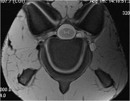
Janes compared 19 Thoroughbreds with CSM to nine control Thoroughbreds; all the horses were an average of about a year old. All animals underwent a neurologic exam and standing cervical radiographs (some in combination with myelograms, which involve injecting a contrast agent into the spinal canal to reveal any compressive lesions on radiographs). After euthanasia, she had MRI studies performed on each horse’s cervical column and evaluations of the images to identify the location, severity, and distribution of bone and cartilage lesions in the articular processes (a particular anatomical structure of the neck vertebrae). Further examinations with micro-CT and histopathology (looking at cell structure under a microscope) were performed on a subset of horses to further characterize the bone and cartilage lesions identified on MRI.
“On average, if you included all lesions, there were significantly more bone and cartilage lesions in our CSM horses as compared to our control horses,” she said. In addition to an increased frequency, she noted increased severity of bone and cartilage lesions. From this study, Janes identified that cervical vertebral lesions were not limited to the compression site in affected animals. This finding brings up the question, “What does the pathology we see say about the pathogenesis (how it came about) of CSM?”
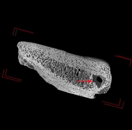
Examining the neck lesions using micro-CT and histology, she found that “all of our superficial lesions, which by definition involved the articular cartilage, fell under the umbrella of osteochondrosis, both in CSM and control horses. Osteochondrosis is a known to occur due to developmental issues in bone growth. Our deep lesions, which involved the bone with no communication to the articular cartilage, were comprised of true bone cysts, osteosclerosis (bone thickening), fibrosis, or cartilage matrix spicules in trabecular bone. Some horses had mixed lesions, which were separate bone and cartilage lesions in the same articular process. Mixed lesions were only identified in the CSM horses, consistent with more severe pathology in diseased horses.”
This is the first report of true bone cysts in the articular processes of horses with CSM. “Bone cysts can occur from developmental issues in skeletal growth,” she said. “The presence of fibrosis is interesting because this could represent a healing response of a previous lesion. Osteosclerosis can occur due to changes in biomechanics on the neck.
“Our bone and cartilage lesions occurred with increased frequency and severity in CSM, regardless of compression site,” she summed up. This suggests more of a generalized issue with vertebral bone and cartilage development in the neck. Further, the types of bone and cartilage lesions observed in the articular processes support the idea that primary developmental abnormalities of the vertebrae with secondary biomechanical change leads to vertebral malformations seen with CSM.