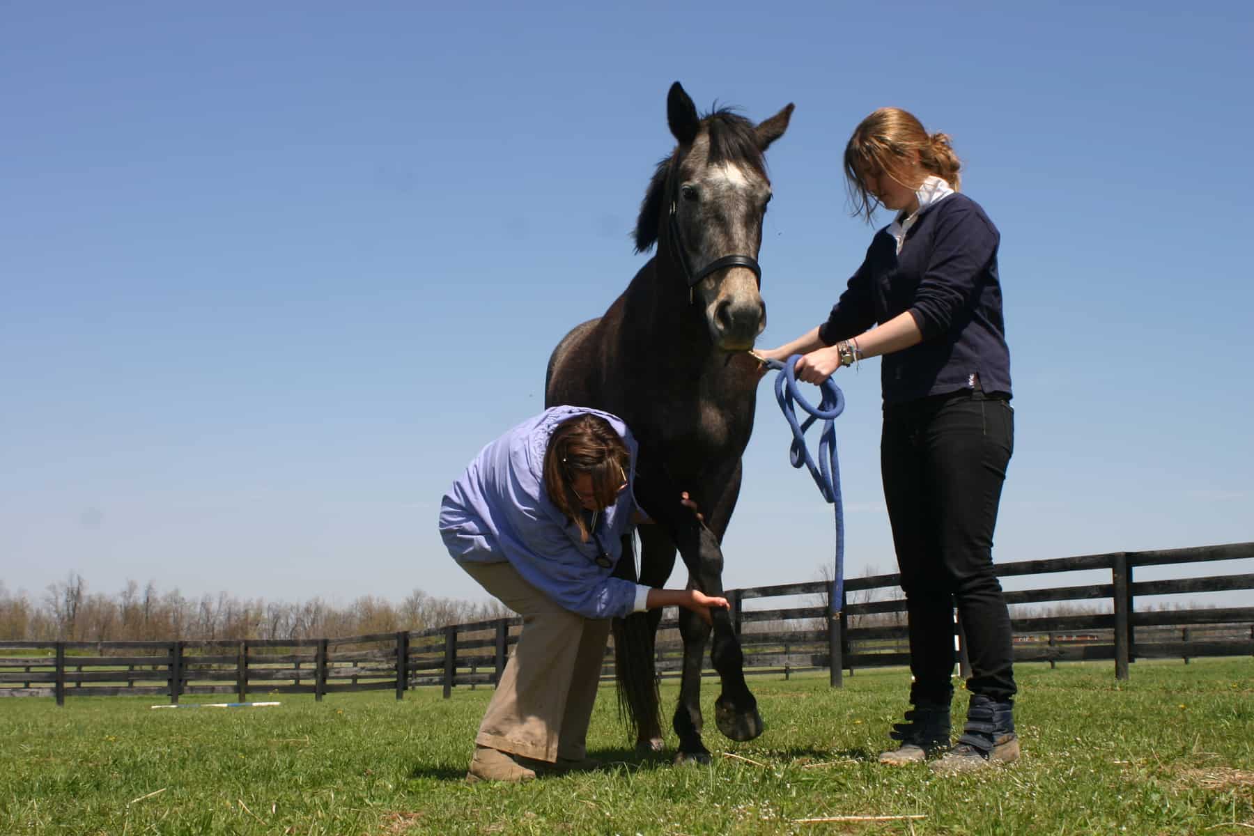
Regardless of the cause, diagnostic evaluation begins with a neurologic examination, which provides anatomic localization of the problem within the central nervous system. Then the diagnostic investigation continues with more focused testing.
If CSM (also known as wobbler syndrome) is strongly suspected, radiographs of the neck should be taken. Narrowing of the cervical vertebral canal in combination with malformation of the cervical vertebrae results in spinal cord compression in CSM patients. Standing lateral radiographs of the cervical vertebrae often reveal bony malformations and probable narrowing of the vertebral canal. Myelography is an important antemortem diagnostic tool and is essential prior to surgical intervention. This condition occurs primarily in young horses (3 months to 1 year of age) where it is a multifactorial disease. In older horses, CSM is often secondary to osteoarthritis of vertebral articular process joints.
The neurologic exam for suspect EPM horses shows asymmetric ataxia, often with upper and lower motor neuron signs and muscle atrophy. Sarcocystis neurona is the most common cause of EPM, but Neospora hughesi infection can also cause similar clinical signs. Several studies of S. neurona demonstrate that horses residing in states with opossums have an exposure rate of 33% to 53%. The exposure rate for N. hughesi appears to be much lower, although less epidemiologic data is available for this organism. Risk factors for S. neurona infection include age (younger than 5 and older than 13 years), time of year (summer and spring more than winter), whether previous cases had been recognized on the farm, presence of a wooded area, and presence of opossums on the farm. Prevalence of the disease was reduced on farms where wildlife had little or no access to feed and if a creek or river was on the premises. Diagnosis of EPM remains a challenge and should begin with physical and neurological examinations. This is followed by measurement of antibodies against the causative organisms in blood and cerebrospinal fluid. Unfortunately, the only definitive test for EPM is a post-mortem examination
Create a free account with TheHorse.com to view this content.
TheHorse.com is home to thousands of free articles about horse health care. In order to access some of our exclusive free content, you must be signed into TheHorse.com.
Start your free account today!
Already have an account?
and continue reading.