A classic rocked-back stance, bounding digital pulses, hooves that are hot to the touch, and clearly painful movement might point to a laminitis diagnosis. But, not all laminitis cases look like this; other symptoms can come into play. Veterinarians must look comprehensively at the clinical picture to not only diagnose the disease but also come up with a treatment plan and prognosis.
At the 2017 American Association of Equine Practitioners Convention, held Nov. 17-22 in San Antonio, Texas, James Belknap, DVM, PhD, Dipl. ACVS, reviewed how to evaluate horses suspected of suffering from laminitis and what information to collect. Belknap is a professor of equine surgery who works on podiatry cases at The Ohio State University College of Veterinary Medicine, in Columbus. He is the editor of Equine Laminitis, the first complete textbook on the disease, published in 2017.
Different Laminitis Types, Same Structural Failure
Laminitis, by definition, is inflammation and possible injury to the hoof’s laminae, which suspend the coffin bone (also called the distal or third phalanx) within the hoof. There are three types of laminitis that have the same general result, Belknap said: structural failure of the laminae (or lamellar failure) that results in coffin bone displacement.
Sepsis-related laminitis usually develops 36 to 48 hours following the onset of sepsis (infection of an organ that results in a systemic inflammatory response). It commonly occurs in horses suffering from pleuropneumonia (bacterial infection of the lung and surrounding pleural cavity), enterocolitis (inflammation/infection of the small and large intestine ), or endometritis (infection of the innermost lining of the uterus, most commonly post-foaling). Sepsis-related laminitis can come on quickly, so as soon as a veterinarian diagnoses one of these conditions, he or she should institute prophylactic (preventive) therapy, said Belknap. The only therapy documented to effectively protect against laminitis in septic horses is cryotherapy (immersing the feet in an ice/water slurry and maintaining them in that state 24 hours a day for several days), he explained.
Supporting-limb laminitis develops in a limb opposite a severely lame limb. Although it is not difficult to identify horses at risk of supporting-limb laminitis, it is difficult to provide prophylactic therapy because the developmental period can be so long; animals commonly do not develop this type of laminitis for weeks to months after the onset of lameness in the opposite limb. Unfortunately, once the animal exhibits signs of supporting-limb laminitis, catastrophic lamellar failure quickly follows.
Endocrinopathic laminitis, on the other hand, has a “slow, insidious” onset, Belknap said. Unlike the catastrophic lamellar failure that leads to crippling lameness in the other two types of laminitis, the laminae appear to fail gradually (over months to years) in endocrinopathic laminitis. Often, the coffin bone undergoes an insidious downward displacement—subtly over time—but the animal does not demonstrate the classic signs of laminitis. The first indication might be something as simple as a bruised sole (due to the slowly displacing coffin bone “pinching” it against the ground surface), and it should be a “rule-out” in any case of bilateral (affecting both feet) fore limb lameness, he said. This is the most common form of laminitis, and the one often associated with equine metabolic syndrome and pituitary pars intermedia dysfunction.
Working Up the Severely Painful Case
Laminitis signs range from mild to severe, but Belknap focused his presentation on assessing the extremely painful patient.
As with any veterinary exam, collecting a thorough history is important. Key factors to note include when the episode began (specifically regarding season and forage availability), any dietary changes, previous lameness issues, and recent infections that could lead to sepsis. The veterinarian should also examine the hoof and surrounding structures for signs of laminitis, such as coronary band separation (from the hoof wall) or solar surface abnormalities.
“The heart rate is extremely important to obtain initially,” Belknap said. “In my view, the heart rate (if not complicated by sepsis, which can result in a substantially increase heart rate) is the most valuable physical parameter regarding assessment of the horse’s duress. Whereas heart rates in the range of 45 to 55 beats per minute are common with moderate pain in laminitis cases, heart rates in the 60s—and especially those in the 70s and 80s—are of great concern not only regarding humane grounds but also prognosis.”
Next, if the horse is able to move, the veterinarian might elect to perform a lameness exam at the walk. Belknap said tests might include walking in a straight line and turning figure-eight patterns on hard and soft surfaces. If endocrinopathic laminitis is the key concern, the veterinarian might have the horse jog on a soft surface to further assess the lameness, depending on its severity.
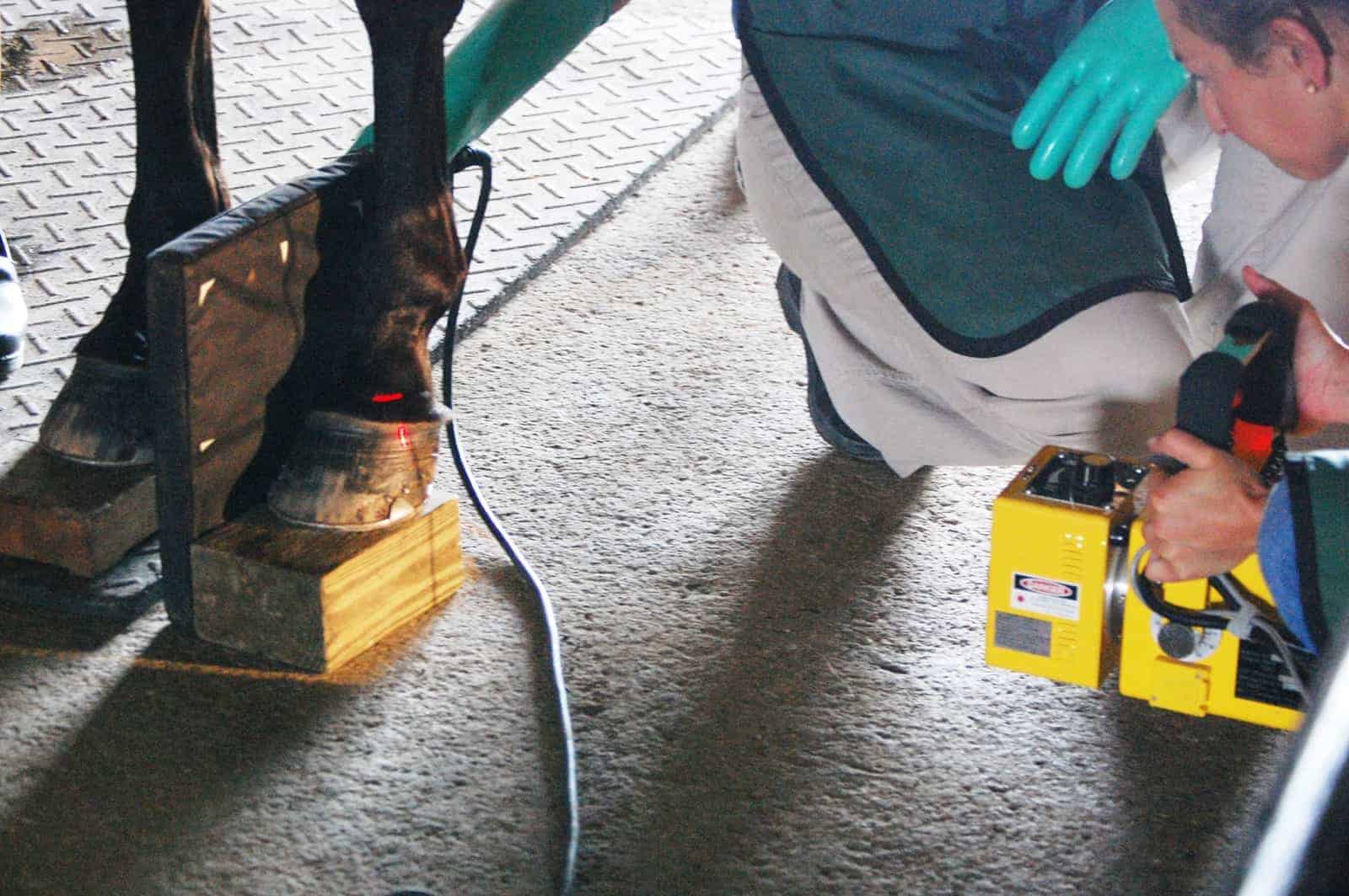
Regardless of which laminitis type a horse has developed, the veterinarian must take steps to determine how much, if any, coffin bone displacement has occurred. For this reason hoof radiographs (X rays) are a must.
Belknap said administering short-lasting nerve blocks in the front feet (the feet most commonly affected) can help rapidly reduce the horse’s pain and allow veterinarians to get better radiographs; they’re able to manipulate the horse’s feet better without making him more painful, which can lead to improved image quality. Blocking the front feet also allows veterinarians to assess any hind-limb involvement. He cautioned that if horses can’t feel their hooves, they could move around too much and cause additional lamellar damage. As such, he recommended attendees add protection and support (such as padded boots) to the hooves to reduce the likelihood of further damage, and he suggested restraining horses until the nerve blocks wear off.
Several different radiographic views can be helpful when assessing laminitic horses, he said:
- Lateral view (from the side)—Veterinarians can see coffin bone rotation (when the tip of the bone points downward), symmetrical sinking (the entire coffin bone displaces downward), and asymmetrical sinking (see sidebar), as well as hoof wall and sole thickness, on this view.
- Dorsal palmar view (front to back)—The most important use of this view is to assess whether the coffin bone is sinking asymmetrically (most commonly a distal, or downward, displacement of the medial, or inner, side of the coffin bone).
Belknap noted that it can be difficult to determine if and how much the coffin bone has moved if baseline radiographs aren’t available. Therefore, he recommended taking serial radiographs to measure ongoing displacement.
In most cases, he said, veterinarians won’t need to collect additional images. The exception might be if a horse is extremely painful but has no evidence of laminitis on radiographs. In these cases veterinarians might have a better chance of seeing lamellar separation on MRI.
Post-Exam Protocols
From there, the practitioner can formulate a treatment plan, as well as a prognosis. Belknap stressed again that serial radiographs are important for monitoring coffin bone displacement (or lack thereof) throughout treatment and recovery.