
Catching and diagnosing equine lameness early, when you first notice something is “not quite right,” gives your horse a better chance for a quick recovery.
Horse owners dread the day their horse “isn’t traveling quite right.” Your gelding might seem a bit off when you’re working him on the longe line. Maybe your mare isn’t striding out like she usually does under saddle.
If your horse is lame, getting a prompt diagnosis might mean the difference between a few days off or a few months.
Early Intervention
“It’s better if you catch it earlier to prevent a more serious layup,” says Sylvia Ouellette, DVM, Dipl. ABVP, of Monrovia, California. “If you can catch the lameness or the problem early, then you can treat it early and get them back to work as opposed to waiting until they’re head- bobbing lame to call.”
Waiting a couple of weeks or months to see if the lameness clears up can make it more difficult to diagnose the problem. Performance horse trainers will sometimes even call the veterinarian when the horse has heat in a leg, but isn’t lame.
“We can palpate a soreness in a ligament, even though the horse isn’t lame, but he has heat there,” says Don Scott Vrono, DVM, of San Dimas, California. “It’s what we call ‘The check-engine light’s on.’ ”
Hands-on: Still the Best
With advanced technologies such as nuclear scintigraphy and MRI, veterinarians can accurately diagnose more lamenesses. Despite all the bells and whistles, though, a lameness diagnosis still begins with an old-fashioned vet call.
Vrono and Ouellette advise owners to keep horses off anti-inflammatories, such as Bute or Banamine, for 24 to 48 hours before the exam because those medications can mask pain and lameness.
“The first thing we do is see how the horse is moving at a walk,” says Vrono. “Then we go over their legs looking for heat or swellings.”
Ouellette likes to look at a horse first without much owner input so she can make an initial objective evaluation. “I will spend the next 20 minutes grilling you on history,” she says, “anything that can help me try to narrow down what I’m seeing.”
The puzzle sometimes is to determine if the problem is actually a lameness. Any information an owner can provide on what led to the situation can be crucial.
“Some lamenesses aren’t really truly lamenesses, but are more performance issues,” says Vrono. Perhaps a horse takes one lead easier than the other, stops at a fence, or won’t pick up his legs over a trail obstacle. Different veterinarians approach distinguishing behavioral issues from lameness issues in different ways, but generally they perform a detailed lameness examination before trying any therapeutic medications.
In addition to watching the horse move at the walk and palpating the legs, a veterinarian will ask to see the horse jog in a straight line and longe in both directions. The practitioner might ask you to jog the horse over multiple surfaces because hard ground can exacerbate bone problems and soft ground can often highlight soft tissue injuries.
Hoof testers will cause a horse to flinch if he feels pain, and this is often an indication of an abscess or hoof bruise.
“I usually start at the bottom and work up,” says Vrono of a lameness examination. “I do a more thorough palpation of the legs, the joints, the tendons, and the ligaments.”
Veterinarians will conduct flexion tests. These range from passive flexion, where the veterinarian flexes a joint to see if it has normal range of motion, to holding a joint in flexion for about a minute and watching the horse jog off. “We look to see if the flexion test exacerbates the lameness,” says Ouellette. “Our job is to try to pinpoint where the pain is coming from that’s causing the lameness.”
Veterinarians will often use the lameness scale adopted by the American Association of Equine Practitioners to, in effect, grade a horse’s lameness. If a horse registers 0, no lameness is perceptible. A 5 is the most extreme (see sidebar).
After the Hands-on Exam
Depending on what the veterinarian has discovered thus far, nerve blocks, radiographs, and/or ultrasound exams are usually next.
Nerve blocks involve injecting a local anesthetic next to the nerves in parts of the leg and seeing if the lame horse jogs off sound. “If the pain is coming from that section you just numbed, the horse will be sound because he can’t feel that pain anymore,” explains Ouellette. “You’ve got to find out where it is before you can find out what it is.”
Ouellette and Vrono advise owners to be prepared to ride their horses during a lameness exam.
“Having a rider on their back is going to make horses behave differently,” Ouellette says, “because they’re now having to compensate for a rider, so they are going to have to move their body a little bit differently. And just that excess weight can exacerbate a lameness.”
What you as a rider can tell a vet will help with the diagnosis. Vrono says, “For example, a dressage rider might be able to feel that a horse is uneven and you might not see it.”
Adds Ouellette, “If you talk to me while you’re riding, you can tell me, ‘Right there. Can you see that?’ “
Looking Inside
A veterinarian might go straight to radiographs or ultrasound, depending on what the flexion tests show. “It depends on how obvious something may seem,” Vrono says. “After we radiograph them, for example, and they have some arthritic changes in their fetlock, sometimes we will go back and block the fetlock separately to see if that’s indeed where the problem is coming from.”
If a veterinarian suspects a hairline fracture, which usually causes a severe lameness, they will want to radiograph before blocking. “If you block it before you X ray it, then those horses can blow their fractures apart,” Vrono explains.
Digital radiographs and ultrasound equipment have revolutionized lameness diagnosis. Veterinarians can examine images immediately instead of having to develop film and return. “If I’m not at the right angle, I can retake that image right then and there,” Ouellette says. Veterinarians also can e-mail digital images to a colleague if they want a second opinion.
Generally, radiographs are ideal for diagnosing bone problems, while ultrasound is better for discovering tendon and ligament injuries. “We do use radiography for suspensory (ligament) injuries,” says Vrono, “because you can look at where the ligament actually attaches to the back of the cannon bone. You can also sometimes see some remodeling on the bone itself where the ligament attaches.”
Advanced Technologies
If the location and cause of the lameness remain elusive, veterinarians might refer your horse to a facility for nuclear scintigraphy, an MRI, or perhaps even a computed tomography (CT) scan. While veterinarian visits, blocks, radiographs, and ultrasounds might each hit your pocketbook in the $200-and-up range, advanced imaging will cost more in the neighborhood of $600 to $2,500. However, these technologies can often help veterinarians find otherwise undetectable lamenesses, and insurance might cover some of these expenses.
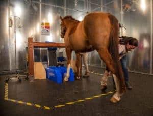
Nuclear scintigraphy, or bone scans, show “hot spots” where bone is actively trying to fix a problem. A more diffuse area is imaged (whole body, including axial skeleton and limbs) than other types of scans, and scintigraphy can provide a “road map” of the horse in its entirety that, in turn, helps direct the course of nerve blocks and localization of lameness. Once the veterinarian localizes the lameness using blocks, he or she will know the exact area to image further.
“A (nuclear scintigraphy) scan can help you find more subtle lamenesses that you can’t really block out,” Vrono says, “sometimes in their back, neck, or upper extremities.”
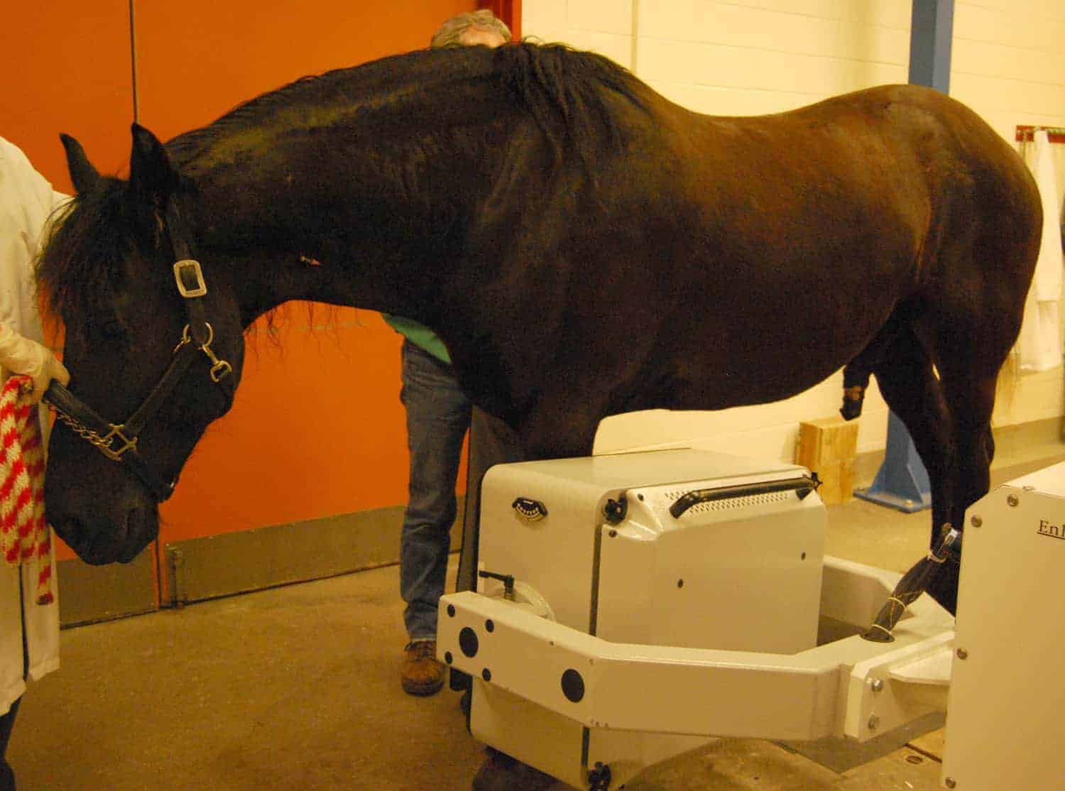
An MRI can yield information about bone and soft tissue, but it is limited to a more focal region than a bone scan (for example, the foot or origin of the suspensory ligament). Many lamenesses stem from the foot, and before MRI technology, many such lamenesses were blamed on navicular disease. Yet the foot includes several major soft tissue structures that can cause lameness, and MRI can often detect the culprit.
“Now that we have MRI, we can actually see what’s going on in the foot,” Ouellette says. “In the past, collateral ligament (injuries) in the foot have been very frustrating. We would get only about 50% of them back (sound after lameness).”
With advanced diagnostics showing her where to focus treatments such as extracorporeal shock wave therapy and IRAP (interleukin-1 receptor antagonist protein), Ouellette says she can now get 90-95% of performance horses back to pre-injury level.
New Diagnostic Technologies
Robin J.W. Bell, BVSc, MVSc, DVCS, MRCVS, formerly of the University of California, Davis, and now of the University of Sydney, in Australia, says sacroiliac disease and high suspensory injuries are among the most difficult lamenesses to diagnose. But new techniques might help.
“We are currently using a purpose-built digital video system to record all our lameness examinations,” says Bell, “allowing for accurate diagnosis after nerve blocks and to help document for rechecks.”
Take-Home Message
Successful lameness diagnosis takes a skilled veterinarian and an owner who can provide a detailed history of how the problem developed. How many and which diagnostic tools will be used depends on where the lameness is located and how severe it is. But with today’s advanced technologies, you have a better chance than ever of finding the problem and starting a treatment regimen that will bring your horse back to soundness.


