Muscle Disease: Immune-Mediated Myopathies
Valberg discussed muscle disease created by immune-mediated situations, describing three possible different manifestations. One type of muscle damage develops subsequent to an outbreak of Streptococcus equi (strangles).
If a horse
- Topics: AAEP Convention, Article, Strangles
Valberg discussed muscle disease created by immune-mediated situations, describing three possible different manifestations. One type of muscle damage develops subsequent to an outbreak of Streptococcus equi (strangles).
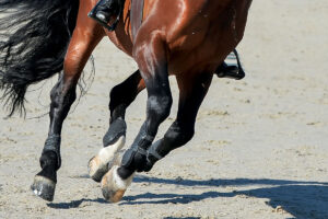
If a horse has strangles, then about three to four weeks later the horse might experience a severe immune-mediated case of muscle damage, also known as S. equi rhabdomyolysis (a form of tying-up). Such a horse might seem fine, then suddenly he develops severe muscle damage and his urine might be a dark coffee color. Most reported cases of this have been in Quarter Horses less than seven years old.
An affected horse has typical signs of a strangles infection: swollen submandibular lymph nodes and/or pus in the guttural pouch caused by S. equi. Then, suddenly the horse demonstrates a stiff gait, firmness of the muscles with severe muscle pain, and eventually he can become recumbent in spite of anti-inflammatory medication and antibiotics. Imminent death in cases of Strep myositis is caused by severe muscle necrosis, subsequent kidney damage, and multiple areas of venous thrombosis (blood clotting in the veins). This is not a result of any specific strain of S. equi, but the inflammatory cascade can produce a toxic shock-like syndrome
A second immune-mediated condition is severe muscle swelling from infarction (localized tissue death resulting from obstruction of the blood supply to the affected site), called infarctive purpura hemorrhagica (PH), that also develops two to four weeks following a respiratory infection associated with S. equi spp.
Deposits of immune complexes of antigen and antibody elicit inflammation in blood vessels (vasculitis) and focal hemorrhages in the muscles. An affected horse becomes stiff and lame, depressed, and he might appear colicky. Other signs of classic PH also appear, such as depression, blood spots on mucous membranes (petechia, or pinpoint purplish red spots, and ecchymoses, which are similar, but larger, spots), and pitting edema of the lower legs, belly, chest, and head.
With the infarctive form of PH, the pectoral, abdominal, and/or hind limb adductor muscles become hard and painful. Infarctions in the bowel will elicit colic and death. In most cases, infarctive PH is rapidly progressive with a high mortality rate; early recognition and aggressive treatment with high doses of corticosteroids and antibiotics might save a horse’s life.
During her presentation, Valberg also discussed a third immune-mediated muscle disease–immune-mediated polymyositis–is characterized by sudden muscle atrophy. In about 40% of these cases, exposure to some respiratory disease or S. equi has been a trigger. This syndrome, so far identified predominantly in Quarter Horses, some Paint horses, an Icelandic horse, a Thoroughbred, and a couple of ponies, is more common than the two previously discussed, and it is very dramatic.
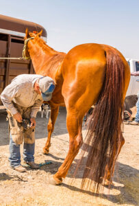
Within three to four days, an affected horse loses all muscle mass, starting on the topline and haunches. Usually the atrophy is symmetrical, but not always. Within a week, more than half of a horse’s muscle mass might waste away, causing dramatic weakness, stiffness, and general malaise. Muscle biopsy of the back or gluteal muscles is important to establish a definitive diagnosis and to rule out PSSM.
Antibiotic treatment is recommended to resolve bacterial respiratory disease, and tapering doses of corticosteroids can limit progression of muscle atrophy. Clinical signs can recur in 40% of cases and might require corticosteroid treatment (although not intramuscularly, since muscle damage has already occurred).
Once the immune-mediated syndrome is interrupted, the muscles should respond and the horse can recover, although in some cases, the full extent of muscle mass might not entirely return to normal.
Get research and health news from the American Association of Equine Practitioners 2006 Convention in The Horse’s AAEP 2006 Wrap-Up sponsored by OCD Equine. Files are available as free PDF downloads
Create a free account with TheHorse.com to view this content.
TheHorse.com is home to thousands of free articles about horse health care. In order to access some of our exclusive free content, you must be signed into TheHorse.com.
Start your free account today!
Already have an account?
and continue reading.

Related Articles


Stay on top of the most recent Horse Health news with