Basic Horse Anatomy and Physiology
The evolution of the horse from a tiny, four-toed animal, perhaps no more than one foot tall, to the variety of equines in existence today, is one of the wonders of nature. During that process of change, the horse evolved over many thousands of years from an animal that predators hunted for food to an animal that became a servant and friend for mankind.
Today’s horses are designed to do one of two things–pull a load with their shoulders or carry riders on their backs. The type of horses utilized for these respective tasks varies a good deal; one is large and ponderous and the other is lighter-boned with less muscle mass. Even within these two types, there are significant differences. For example, the conformation of a roping or cutting horse is different from that of the American Saddlebred. Yet there is a basic sameness to anatomy.
In this opening article of our 12-part series on equine anatomy and physiology, we will attempt to paint a picture of how today’s horse is constructed and what this means in the realm of form to function. We will not be quoting a lot of sources, for the most part, because the information to be presented is an amalgamation of what has been recorded in thousands of scientific papers, textbooks, and manuals as knowledge has been gained and disseminated through the years. However, we would be remiss in not calling attention to two valuable sources upon which we shall draw heavily. Both are written at the layman level. They are Horses and Horsemanship by the late M.E. Ensminger, BS, MA, PhD, and The Coloring Atlas of Horse Anatomy by Robert A. Kainer, DVM, MS, and Thomas O. McCracken, MS, both of whom were at Colorado State University.
McCracken, who resigned from Colorado State in 1994 to enter private business, has authored seven books, and today he is president of Biographix, LLC. Early in his career, McCracken spent two years in Saudi Arabia as chief medical illustrator for the King Faisal Specialist Hospital and Research Center in Riyadh. Through the years, he has received many honors and awards of excellence for his work.
Kainer joined the anatomy faculty at Colorado State in 1961 and remained there for 27 years. He, too, has won a variety of awards and honors for his work.
Needless to say, since there are entire books written on equid anatomy and physiology, it would be impossible to address the entire animal in one article. Therefore, this month we look in-depth at the horse’s largest organ, its skin, and details of the nomenclature used to discuss anatomy and physiology, as well as lightly touch on structures that will be covered in-depth in the coming year. In the months ahead we’ll have articles on joints, the foreleg, the hind limb, the hoof, the head and neck, the back, muscles, tendons and ligaments, and the digestive, circulatory, respiratory, and reproductive systems.
A Glossary to Get You Started
We will begin this discussion of equine anatomy with Kainer’s and McCracken’s explanation of terminology that is routine for veterinarians and researchers, but can be confusing to some horse owners. Along with the two authors’ terminology, we’ll add examples to demonstrate how the terminology might be employed. Understanding the terminology is essential when discussing various conditions with a veterinarian.
Here is some terminology, accompanied by illustrations, to help you visualize:
On Side–The horse’s left side; also called near side. Example: Normally, the side on which we mount and dismount.
Off Side–Right side of the horse; also called far side. Example: One theory of why we mount from the left instead of the right is that warriors and soldiers of old generally carried a sword on the left side and this would get in the way if swinging up from the right or off side of the horse.
Dorsal–Parts of the horse’s anatomy toward his back (dorsum). Example: The point of the croup is dorsal to the stifle.
Ventral–Anatomy toward the belly (venter). Example: The stallion’s reproductive organs are ventral to his flank.
Cranial–That part of the horse’s structure above the knees and hocks located closer to the skull (cranium). Example: The withers are cranial to the tail.
Caudal–That part of the horse’s structure above the knees and hocks located closer to the tail (cauda). Example: The horse’s back is caudal to his neck.
Rostral–That part of the structure located closer to the nose (rostrum). Example: The eyes are rostral to the ears.
Proximal–A location toward the attached end of a limb. Example: The proximal end of the cannon bone connects with the knee.
Distal–Indicates a location toward the free end of a limb–the part that is farther away from the body. Example: The distal end of the cannon bone connects with the long pastern bone or third phalanx.
Median Plane–This divides the horse’s body into right and left halves (median means in the middle).
Sagittal Plane–Any plane parallel to the median plane. Example: A plane dividing the right and left sides of a hoof.
Medial–Structures located closer to the median plane. Example: When you look at a horse’s left side, you see the medial surfaces of his right limbs.
Lateral–Structures located away from the median plane. Example: When you look at a horse’s left side, you see the lateral surfaces of his left limbs.
Transverse Plane–Passes through the head, trunk, or limb perpendicular to the part’s long axis. Example: An ultrasound image gives a transverse view of the limb.
Dorsal Plane–Passes through a body part parallel to its dorsal surfaces.
There are subtle shifts in terminology as we move about the equine anatomy. For example, the two writers point out, “Distal to and including the carpus (knee), dorsal replaces cranial and palmar replaces caudal. Distal to and including the hock, dorsal replaces cranial, but plantar replaces caudal.” This means that when you are talking about the lower limbs, you use the word dorsal to mean frontward instead of cranial, and instead of using caudal to mean rearward, you use palmar (for the front limb) or plantar (for the hind limb).
The solar surface of the hoof is the part that contacts the ground.
When looking at a horse’s limb from the front, an axial structure is located toward the axis (center of the limb) and an abaxial structure is located away from the axis.
Classifying the Horse
Before leaving our Atlas authors, we’ll pass along their explanation of proper terminology for this animal we are discussing, along with what they consider to be identifying characteristics:
“Horses and their close relatives, donkeys and zebras, are in the mammalian order of odd-toed, hoofed animals (Perissodactyla) as are their distant relatives, rhinoceroses and tapirs. The horse, Equus caballus, is an equid, a member of the horse family Equidae. The adjective equine is frequently used improperly as a noun.”
They point out that characteristics of equids include:
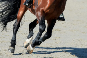
1) Highly specialized limbs, each with one digit (the third) and with the main muscle mass of the limb situated close to the body’s trunk.
2) Large paranasal sinuses within the skull.
3) Guttural pouches, which are large outpocketings of the auditory tubes that extend from the nasopharynx to the middle ears.
4) High-crowned permanent teeth that grow for a long time–a feature used to determine the age of a horse.
5) A simple stomach followed by a long small intestine and a large, complicated large intestine where fermentation of feed occurs.
6) Well-developed skin glands.
7) Large heart and lungs.
8) A uterus with short horns and a relatively large body, and a prominent depression in each ovary where the egg cells are released.
9) A large, vascular penis and a complete set of male accessory sex glands.
There are similarities between human and equine anatomy, but there are many differences too. For example, the human knee and the equine stifle have a lot in common. The equine radius is similar to the human forearm. Equine carpal (knee) bones compare to human wrist bones. And equine splint bones compare to human fingers.
There is also a similarity in the cooling process during exercise–both humans and horses cool their bodies via sweating. Yet, the two are worlds apart when looking at the digestive process.
Skin
When discussing equine anatomy, what better place to start than with the skin, the horse’s largest organ, ranging from 12-24% of the animal’s total weight depending on age. It serves a number of functions. It protects underlying tissues from injury, drying, water absorption, and bacterial invasion. Another important role involves thermoregulation (regulation of body temperature). The skin also excretes water and salts through sweat glands, senses the environment, and synthesizes vitamin D in response to sunlight.
The skin consists of various cellular and tissue components. There are two layers, the epidermis and the dermis (subcutis), with the epidermis being the outer layer. The two are attached by collagenous and elastic connective tissue.
The main activity of the epidermis is to produce two types of protein–keratin and melanin. Keratin, the principal component of the epidermis, is a simple protein characterized by its insolubility (it won’t dissolve) and fibrous structure. It serves a supportive and protective function, including the shedding of water. Melanin is the dark, shapeless pigment of the skin and hair.
Another cell type in the epidermis is the Langerhans’ cell, a non-neural cell that is active in the immune response and possibly in the regulation of keratin formation.
The most important part of the epidermis is the superficial layer–known as the stratum corneum–since much of the functional activity of the skin resides here. The proper functioning of this superficial layer is dependent on the structural arrangement of the keratin it contains and on its lipids–fats and fat-like substances characterized by being water-insoluble. Lipids and keratin combine to waterproof the skin and prevent various agents from entering the body. If this superficial layer of the epidermis were removed, the skin would be like a mucous membrane that is easily permeated by water and a variety of agents.
The hair follicles in a horse’s skin are quite simple, with a single hair emerging from each pore. The hair that emerges from these pores serves as frontline protection for the horse. First, a heavier growth in cold weather helps to provide warmth and a lighter coat during summer facilitates the cooling process. The hair also serves as an important filtering system. For example, ultraviolet light is filtered by the hair coat and is absorbed by melanin granules in the epidermis and hair. Hair is composed of keratinized epithelial cells.
Each hair consists of a shaft and a root that is contained in a depression known as the hair follicle. Associated with hair follicles are sebaceous (oil) glands and bundles of smooth muscle connecting the side of the hair to the dermis. The sebaceous glands secrete an oily substance into the follicles and thus to the epidermal surface. The oil lubricates the skin to prevent excessive evaporation that can result in dry skin.
There are two types of sweat glands in the horse–the apocrine and the eccrine glands. The apocrine glands are spread throughout the skin, while the eccrine glands are found only in the frog of the hoof and, thus, play a very limited role in the cooling process. The horse sweats the most of any of the domestic species. The sweat glands are simple, coiled, tubular glands that open independently of the hair follicle.
Nerve endings in the dermis, just under the epidermis, are called sensory nerves and carry sensations of pressure, pain, heat, and cold. In contrast, motor nerves cause the sweat glands to secrete.
When sweat reaches the surface of the skin, it evaporates. This has a cooling effect on the horse’s body. Since sweating is the horse’s primary method of cooling the body during exercise, cooling can be severely compromised when evaporation of that sweat does not take place, such as on a hot, humid day. Unfortunately, nature didn’t provide much in the way of backup measures for hot, humid weather, and it is up to the humans involved to help out by providing plenty of water to drink and cool mists or baths to cool the body when horses must be exercised in such conditions.
Research has shown that a horse’s body temperature can rise to a dangerous level very quickly when adequate cooling does not occur. Without proper cooling, body temperatures during endurance rides, for example, can rise to 106ºF (normal temperature is 99.5-101.5ºF), and the ultimate result can be heat stroke.
There is more involved in the thermoregulation department than just sweating. An intricate system of blood vessels must first carry heat from the core of the body to the skin so that it can be dissipated via sweating. When heat buildup overpowers the horse’s ability to dissipate it, the result can be deadly.
The problem develops something likes this: As heat accumulates, blood flow to the skin is increased to speed up the transportation of heat from the core of the body to the skin surface. As exercise increases, however, sweat loss leads to dehydration and the loss of plasma water from the bloodstream. This results in a decrease in the volume of blood that is carrying heat to the surface. The result is an increased heart rate as the horse’s body desperately tries to make up for the decreased volume of blood. Very quickly, body temperature rises and extreme fatigue sets in.
A problem that afflicts some horses and compromises the thermoregulatory system is anhidrosis or dry coat. Horses with this affliction simply can’t sweat. It appears that anhidrosis is climate-specific; the signs are found in areas with a hot, humid climate, such as Florida during the summer.
No one is sure what causes anhidrosis, and there is no effective way to treat it other than to move affected horses to a cooler climate. Often, racehorses that suffer from anhidrosis in Florida will show no signs of the affliction when moved to the cooler Midwest.
The growth and shedding of hair coats follows a definite cyclical pattern. A heavier coat grows when days shorten and weather turns colder, with shorter periods of daylight, and is shed when days become longer and warmer and there are longer periods of light.
Again, we turn to McCracken and Kainer for an explanation of what occurs: “Horses shed hair mostly in the spring, beginning with the belly and sides and continuing up onto the back. The hair cycle includes an actively growing stage (anagen) followed by a stage (catagen) during which the hair matrix sort of shrinks (atrophies) and peels away from its blood supply. A longer, quiescent period (telogen) then occurs in which the hair (club hair) separates from the hair matrix, but remains in the follicle. The matrix later becomes active and begins to grow a new hair that pushes out the old club hair (shedding).”
There are health conditions that can overpower the forces of nature in the shedding department. For example, horses suffering from Cushing’s disease often carry a heavy coat even during long-light, hot summer days.
The color of a horse’s hair is determined by a rather complicated genetic process involving the amount of pigment in the hair. There are three basic hair pigments–black, brown, and yellow. The great variation in color has to do with combinations or absences of these three pigments.
Skin Problems
The skin, as the first line of defense against the elements, insects, and antigens, is under almost constant attack from one source or another. In many cases, the conditions are not life-threatening, but produce a great deal of irritation to the horse and can lead to more serious consequences.
Here are some of the attackers that make war on the skin:
Dermatitis–This is an inclusive term for inflammation of the skin that can be produced by many agents, such as external irritants; burns; allergens; trauma; bacteria; viral, parasitic or fungal infections; and systemic diseases. When dermatitis surfaces, the important thing is to determine the underlying cause, as treatment can vary a great deal. An example of dermatitis is ringworm, which is actually an infection of keratinized tissue by one of several types of fungi. The most common form of ringworm–which is easily transmitted from animals to humans–produces skin lesions that appear as small rounded spots in the skin. The lesions eventually form blisters and break, leaving scabs in their wake.
Rain Rot–This condition carries the scientific name of dermatophilosis. It is a form of dermatitis that is more common in wet, humid climates than in arid areas. The condition is caused by a fungus that is capable of creating infection when conditions are right, such as during hot and humid periods. The infection results in scabby spots that are devoid of hair when rubbed free.
Mange–This condition is caused by several species of mites. Mange is characterized by loss of hair, itching, and intense irritation to the skin.
Lice–Horses can harbor two species of lice. One species, Damalinia equi, is the horse biting louse and the other, Haematopinus asini, is the horse sucking louse. Afflicted horses will show signs of itching by constantly rubbing and biting at affected areas. Lice are most often found in unthrifty horses and quickly add to the poor condition status. In severe infestations, the horse might lose patches of hair.
Dandruff–This is a condition where the horse’s skin becomes scaly and the hair coat often is dry and dirty. In some instances, the hair will fall out. Grooming can help to stimulate the sebaceous glands; exercise and a proper diet can prevent this problem and can work to solve it if it does occur.
Biting Insects–During the summer months, horses are under almost constant attack by one type of biting insect or another, including flies, mosquitoes, and ticks. Not only are these pests irritating to the horse, but they also spread diseases such as West Nile virus, equine infectious anemia, and encephalitis. Responsible horse owners should take all steps that are possible to prevent these disease-carrying pests from attacking the animals in their charge.
Hyperelastosis cutis (HC)–This is an inherited condition of Quarter Horses from a particular bloodline. Earlier, we mentioned that the epidermis and dermis are held together by collagenous material, sort of like glue that can bind two substances. When a horse inherits the genes that cause HC, the collagenous material is defective and unable to hold the two skin layers together; the result is tearing of the skin, which often results in a death sentence for the horse. There is no known treatment.
Stay Tuned…
There, in short form, is a description of the equine skin and its functions. In upcoming articles, we will concentrate on different parts of the horse’s anatomy with an in-depth look at how they are structured, the functions they serve, and what is involved in keeping them healthy.
Here is a brief look at those upcoming article topics, to whet your appetite.
The equine skull houses the brain and the most important organs of sense. There are 34 bones in the equine skull, most of them flat. During the birthing process, these bones yield and overlap, allowing the skull to be somewhat compressed and thus allowing for easier parturition.
The horse has unique eyes–they are both monocular and binocular. Monocular means that the horse can see objects with one eye. As a result, the brain often gets two images simultaneously. With binocular vision the horse can focus with both eyes and the brain is receiving only one signal. Humans have binocular vision only.
From the equine head, we move to the neck. There are seven cervical vertebrae in the horse’s neck, with the spinal cord running through them. The neck blends into the back at the withers.
We come now to the horse’s rib structure. Normally, there are 18 pairs of ribs in the horse, but a nineteenth rib on one side or both is not considered all that rare. Eight pairs of ribs are known as true ribs. They join the segments of the sternum or breastbone. The other 10 are known as floating ribs–merely overlapping and being attached to each other. The seventh and eighth ribs are the longest. Those toward the rear are much shorter.
Next we switch to the horse’s thoracic or front limbs. Each of these consists of the scapula, humerus, radius, ulna, seven or eight carpal bones, cannon bone, two splint bones, two proximal sesamoid bones, large pastern bone, small pastern bone, navicular bone, and coffin bone. The structure of these bones and their functional relationship to each other helps determines a horse’s athletic potential and its capability of remaining sound even under an exacting exercise and performance regimen.
The angles at which the bones connect have much to do with the horse’s physical capability and its continued well being.
The leg bones, as well as other bones, are connected at joints. There are three types of equine joints–synovial, which are movable; cartilaginous, which are slightly movable; and fibrous, which are immovable. The joints that connect the horses’ front leg bones are synovial. A synovial joint consists of two bone ends, covered by articular cartilage. The cartilage within the joint is smooth and resilient, allowing for frictionless movement. Ligaments keep the bones in proper support and relationship.
In the rear legs, we’ll start at the spine where the ilium, the largest of three bones in the pelvis, connects with the spinal column. The ilium angles down and rearward to blend into the femur or thigh bone. The femur angles slightly forward to the stifle. Beginning at the stifle (comparable to the knee in humans) is the tibia, which continues downward to the hock. From the hock to the pastern is the metatarsus or rear cannon bone, which connects with the long pastern bone; next is the short pastern bone, and lastly the coffin bone. Both front and rear limbs are connected and supported by ligaments, tendons, and muscles that provide attachment and mobility.
Nature didn’t do the horse many favors when designing the way in which it processes food. Its stomach can hold only small amounts of food–eight to 16 quarts at a time. The stomach empties into the small intestine, a tube that connects with the large intestine. The small intestine is approximately 70 feet long, with a capacity of about 12 gallons. The large intestine of the horse is divided into four parts–the cecum, which is about four feet long and one foot wide; the great colon, which is about 12 feet long and 10 inches wide; the small colon, which is about 10 feet long and four inches wide; and the rectum.
In the reproductive department, the main sex organs of the mare are two ovaries that produce eggs, which are housed in follicles. When an egg is mature, it leaves the follicle and travels through the infundibulum and oviduct to the uterus. If fertilized, it grows within the uterus from a minute structure into a foal that is born through the vagina.
The primary sex organs of the stallion are the two testicles, which produce semen and testosterone, and the penis.
A horse’s respiratory system is designed–as is the case with other mammals–to exchange oxygen and carbon dioxide. Air enters the nostrils, passes through the long nasal cavity over the pharynx and larynx, then enters the trachea that carries it to the lungs.
A horse’s cardiovascular system consists of blood, a series of blood vessels through which blood flows, and a heart that pumps blood throughout the horse’s body. Blood is comprised of red blood cells, white blood cells, and platelets. The red blood cells have a protein known as hemoglobin that binds oxygen and conveys it to the muscles. The white blood cells function in the immune system by defending against foreign invaders, such as harmful bacteria, and the platelets are involved with blood clotting.
Next month, we’ll start with an in-depth look at joints.

Related Articles


Stay on top of the most recent Horse Health news with