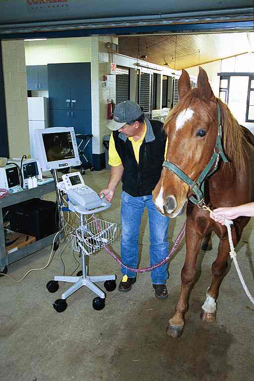
At the 2012 American Association of Equine Practitioners convention, held Dec. 1-5 in Anaheim, Calif., Michelle Henry Barton, DVM, PhD, Dipl. ACVIM, Fuller E. Callaway Endowed Chair and professor of Large Animal Medicine at the University of Georgia’s College of Veterinary Medicine, reviewed transabdominal ultrasound techniques for diagnosing colic caused by problems in the small intestine. Veterinarians can use a low-frequency, curvilinear ultrasound probe placed on the horse’s flank to assess key features of the small intestine, including motility, degree of distension, intestinal wall thickness, and intestinal contents.
Barton said veterinarians can identify normal small intestine on transabdominal ultrasound in only 10-30% of healthy, fed horses. In the healthy horse, waves of contraction called peristalsis keep ingested food moving along the intestine. When this normal motility gets interrupted (either by intestinal paralysis or by mechanical obstruction such as an impaction or a twist in the bowel), it’s called ileus. Veterinarians can identify the distinct pattern of ileus using ultrasonography. As gas builds up in the intestine, it creates an increasing series of hairpin turns that make the intestine resemble a series of switchback trails rather than a long, winding road.
In mild cases of ileus resulting from paralysis secondary to a medical condition such as enteritis (inflammation of the small intestine) the intestinal wall might appear normal, whereas in more severe cases it might look thicker and possess irregular “lasagna noodle” mucosa. In either case, this type of ileus should resolve gradually with fluid therapy and time, said Barton, and veterinarians should see some motility on ultrasound.
In contrast, mechanical ileus continues to worsen, despite therapy, resulting in a larger number of hairpin turns. In some cases the intestine becomes so distended that the loops of bowel look like nearly perfect circles when the probe is turned so as to view a cross-section of the intestine.
Veterinarians can also use ultrasound to distinguish between strangulating and nonstrangulating obstructive lesions. In the case of nonstrangulating obstructions, ultrasound might help the veterinarian determine if the obstruction is severe enough to warrant surgery.
In summary, Barton said ultrasound can be a valuable aid to practitioners when assessing the cause and severity of equine colic secondary to disease of the small intestine.