
How to Take Foot Radiographs (AAEP 2008)
- Topics: Article
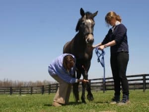
Taking radiographs (X rays) of horses' feet is "arguably the most common form of imaging performed by veterinarians–for lameness, prepurchase, laminitis, and podiatry examinations," notes Keith Merritt, DVM, owner of Merritt & Associates Equine Hospital in Wauconda, Ill. He presented a discussion of how to properly take foot radiographs at the 2008 American Association of Equine Practitioners convention, held Dec. 6-10 in San Diego, Calif.
Radiographs provide information for making diagnoses, planning treatments, and guiding trimming and shoeing. "The quality of the radiographs and the final product generated are dependent on the preparation of the foot, the position of the foot, and the views required for a particular study," he noted.
He offered the following tips on preparing the foot for radiographs:
-
The foot (from the fetlock down) should be thoroughly cleaned, the sole should be wire-brushed, the frog sulci should be picked and trimmed, exfoliating sole should be trimmed out, and the sulci should be packed to eliminate gas shadows. "A properly prepared foot eliminates artifacts (misleading imaging) that can lead to misdiagnosis," he advised.
-
For a laminitis exam, the wall must be marked with a radiopaque substance (such as barium paste) from the coronary band to the end of the hoof wall.
-
Merritt recommends carefully removing shoes for purchase examination, as clips, bars, and nails can hide areas of interest in the foot. However, for a podiatry study it is useful to radiograph the feet with the shoes on to see the position of the shoe relative to internal structures. If shoes are removed and not replaced, the foot should be protected with impression material, cotton, etc., until the shoes can be reset.
Positioning the feet and equipment also requires attention to detail. "Any misalignment, either with the foot not being positioned properly or the radiograph capturing plate or the X ray machine not being aligned, will cause distortion of the bones of the digit (on the radiograph image)," Merritt warned. "This can lead to a misdiagnosis or the wrong information being provided to the farrier
Create a free account with TheHorse.com to view this content.
TheHorse.com is home to thousands of free articles about horse health care. In order to access some of our exclusive free content, you must be signed into TheHorse.com.
Start your free account today!
Already have an account?
and continue reading.
Written by:
Christy M. West
Related Articles


Stay on top of the most recent Horse Health news with


