Don’t jump to trotting lame horses and blocking them bottom to top to search for a diagnosis, and don’t overinterpret imaging findings that might not truly and fully explain the horse’s lameness. This was one of many take-home messages—and perhaps the most important—Jean-Marie Denoix, DVM, PhD, Dipl. ACVSMR, ECVSMR, relayed during his presentation at the 2021 American Association of Equine Practitioners (AAEP) Convention, held Dec. 4-8, in Nashville, Tennessee.
The AAEP invited Denoix, who is founder and past director of ENVA’s Centre d’Imagerie et de Recherche sur les Affections Locomotrices Equines, in France, to deliver the prestigious State of the Art Lecture based on his extensive experience and commitment to understanding equine lameness over the course of his career. Denoix explained that the goal of his presentation was “to improve our information on clinical manifestations of lameness and to better interpret and understand them.” He suggested practitioners use this information to reach a diagnosis with fewer blocks—regional anesthesia involving nerve and joint blocks to desensitize specific anatomic structures in the limbs.
In fact, Denoix said, in many cases a careful clinical examination will reveal all the information practitioners need to reach a diagnosis.
“Sometimes there is no need to block at all, and you can save time,” he said.
To perform a complete clinical examination, Denoix recommended practitioners examine the horse in the following scenarios:
- Walking and trotting in hand on a firm surface;
- Walking straight, and trotting straight, right and left circles, and a figure eight;
- Moving on soft surfaces;
- Cantering left and right circles; and
- At work (this latter condition being essential, he said).
Slow Is Steady and Steady Is Fast
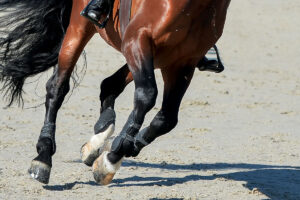
While Denoix addressed each component of the clinical examination, an important part of his presentation focused on examining horses at the walk.
Why so much emphasis on the walk?
“You often get more specific information about the lameness than (with) the other gaits,” said Denoix. “It can be a big mistake clinically to jump too quickly to the trot and blocks.”
Demonstrating his point and relaying the importance of addressing functional anatomy and biomechanics during a lameness examination, Denoix shared a series of cases. Here are some examples of how Denoix uses functional anatomy to diagnose lameness at the walk:
Case 1
The horse presented with a toe-touching lameness of the right front limb and did not want to extend the fetlock during the non-weight-bearing swing phase of the stride. Lameness was localized to the proximal check ligament (accessory ligament of the superficial flexor tendon). However, based on Denoix’s clinical experience, he knew the horse was too lame to have simple check ligament disease alone. Further examination and diagnostic imaging revealed an osteochondroma (cartilage and bone overgrowth) in contact with the proximal check ligament.
“The osteochondroma more clearly explained why the horse is lame and doesn’t want to extend the fetlock,” Denoix said. “Reduced fetlock extension reduces the interference between the check ligament and osteochondroma.”
Case 2
This horse also presented with a tremendous reduction in the cranial phase of the stride and profound lack of fetlock extension. This horse, however, was diagnosed with a fracture at the base of the medial proximal sesamoid bone, which is where the oblique sesamoidean ligament originates.
“Why is this horse reducing cranial phase and reducing fetlock extension?” Denoix asked. “Because peak of load of the suspensory ligament occurs during cranial phase of the stride
and fetlock extension creating pain on the fracture is limited during the caudal phase of the stride by the tension of the deep digital flexor tendon.”
Case 3
This case showed a horse, again at the walk in a straight line on a hard surface, reducing the cranial portion of the swing phase, meaning he had trouble moving the leg forward in contrast to the horse in Case 2, reducing the cranial phase of the stance phase of the gait.
“This horse is unable to protract the leg even when the leg is not in contact with the ground surface while there is no load on the leg,” Denoix explained.
This was a key take-home Denoix emphasized—that practitioners must look at not only the stance phase but also the swing phase.
He diagnosed this horse with osteochondrodysplasia of the shoulder. From a functional anatomy standpoint, the horse is lame because the supraspinatus muscle contraction induces pressure over the scapulohumeral joint, so the horse reduces the swing phase to reduce that pressure.
“Pain and mechanical limitation induced by the lesion causes this lameness,” said Denoix.
At this point in the presentation, it was clear that to diagnose lameness using functional anatomy, practitioners must have a commanding knowledge it. Denoix highlighted this in the following case:
Case 4
He showed a video of a horse with what he described as a tremendous reduction in the cranial swing phase of the left front.
“This horse was blocked everywhere, and we finally radiographed the neck, finding osteochondral fragmentation between C6 and C7, as well as chronic synovitis,” he explained. “Ultrasonography confirmed the findings.”
Despite having a diagnosis, Denoix again took the lameness examination one step further, asking, “Why was there such a reduction in swing phase?”
In this case the lesion resulted in entrapment of the C7 nerve, a major root of the brachial plexus. This nerve provides the main part of the suprascapular nerve, which innervates the supraspinatus muscle. Thus, nerve degeneration of this root explains the reduction in the cranial phase of the stride.
This case is an example of a condition that Denoix called cervicobrachial (relating to the neck and arm) syndrome. He explained that several diseases affecting the brachial plexus can induce changes in the neurophysiology of the nerves in that region, all resulting in a decreased cranial phase of the stride.
Case 5
As another example of the importance of examining horses at the walk, Denoix presented the case of a horse with a reduced caudal phase of the stride with lack of fetlock extension. This horse had a broken shoe, and radiographs revealed a fracture of the palmar process of the third phalanx (P3).
“How do you explain how this fracture is responsible for the reduced caudal phase of the stride?” Denoix queried. “Because the deep digital flexor tendon (DDFT) has a wide insertion involving the palmar process of P3, and in this case the DDFT crosses the fracture line. The tension of the DDFT is higher during the caudal phase of the stride, so the mare wants to reduce that tension.”
This case also supported Denoix’s mantra that “there must be good correlation between clinical manifestation and imaging.”
Why Walk When You Can Run?
“The walk is a pedagogical gait and is very informative,” said Denoix. “The clinical expression of the lameness is superior to what can be seen at a trot or canter. This is because there is increased contact between the foot and the ground, so the caudal phase defects are highlighted, as well as the cranial phase due to slower protraction.”
At the walk, he said, there is little activation of the passive mechanisms, which means the kinematics and displacement of the joints induce more clinical manifestations than the load itself (i.e., stance-phase lameness).
For a complete examination, however, fully assess each horse at each gait on different surfaces and while at work. Denoix’s presentation, available online for conference attendees, provides a plethora of real-world examples that delves deeper into the “why” of equine lameness.